News Story
New Microscopy Technique Sheds Light on How Cells Sense Environment
Dr. Giuliano Scarcelli
Fischell Department of Bioengineering (BIOE) assistant professor Giuliano Scarcelli is collaborating with researchers from the United Kingdom who have developed a new microscopy method that could revolutionize scientists’ understanding of how cells find their way through the body.
The research, published this month in Nature Cell Biology, is led by Malte Gather, a professor with the School of Physics and Astronomy at the University of St. Andrews, United Kingdom, and a long-time collaborator with Scarcelli and fellow researchers in the fields of optics and biophysics.
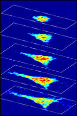
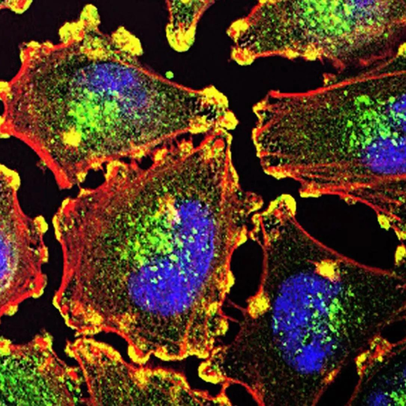
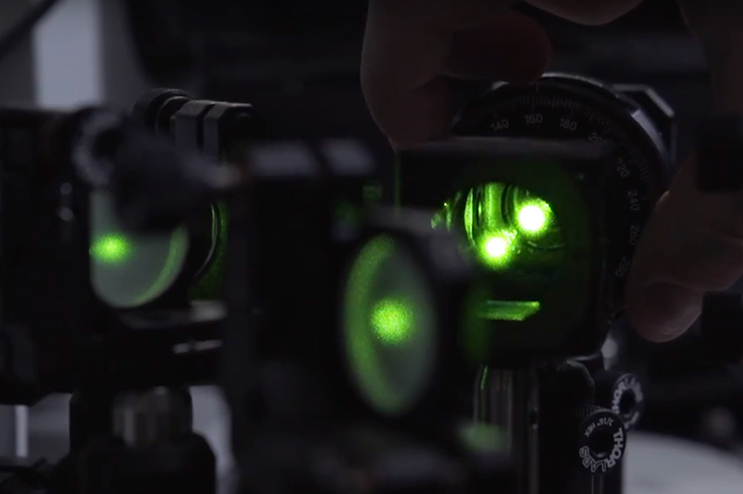
The new microscopy method, Elastic Resonator Interference Stress Microscopy (ERISM), images the extremely weak mechanical forces that living cells apply when they move, divide, and probe their environment. Forces exerted by cells are fundamental for many physiological processes including locomotion – such as during immune response or tumor metastasis – cell growth, wound healing, and tissue formation and repair. As such, researchers have long sought a method for monitoring these forces in a robust and non-disruptive manner.
“Our microscopy records very high color resolution images of the light reflected by a thin and soft probe,” Gather said. “From these images, we then create a highly accurate map of the thickness of the probe – with mind-blowing precision of one-billionth part of a meter. If cells apply forces to the probe, the probe thickness changes locally, thus providing information about the position and magnitude of the applied forces. Although researchers have recorded forces applied by cells before, our interface-based approach gives an unprecedented resolution and, in addition, provides an internal reference that makes our technique extremely robust and relatively easy to use.”
According to a recent press release issued by St. Andrews, this robustness means that measuring cell forces could soon become a tool in clinical diagnostics, through which doctors could complement existing techniques to assess the invasiveness of cancer.
“Cells are not only biochemical entities as one might think,” Scarcelli said. “They are also little physical machines. But, while we have a full arsenal of techniques to study molecular pathways, tools to characterize physical interactions are scarce and inherently more difficult to build. This force sensor is so accurate and fast that it could open a new dimension in how we understand cellular processes.”
Scarcelli’s work with the University of St. Andrews represents another step in his efforts to advance understanding of some of the body’s most complex and spectacular processes, such as those relating to the central nervous system (CNS) or cancer metastasis. Scarcelli’s collaboration on the ERISM project is part of his 2014 Human Frontier Science Program (HFSP) Young Investigator award. The three-year, $1,050,000 grant is titled Neurons feel the force: new photonic tools to unravel the development of the nervous system, and featured the development of novel microscopy techniques to further understanding of the CNS. The award fostered Scarcelli’s collaborations with Gather and fellow co-author on the Nature Cell Biology paper Kristian Franze (Department of Physiology, Development and Neuroscience, University of Cambridge, United Kingdom).
In addition to Gather, Franze, and Scarcelli, the following researchers served as co-authors on the paper: Nils M. Kronenberg, Philipp Liehm, and Anja Steude, (School of Physics and Astronomy, University of St. Andrews); Johanna A. Knipper (Institute of Immunology and Infection Research, University of Edinburgh); Jessica G. Borger (Institute of Immunology and Infection Research, University of Edinburgh); and Simon J. Powis (School of Medicine, University of St. Andrews).
The full paper, Long-term imaging of cellular forces with high precision by elastic interference stress microscopy, is accessible online.
Published June 29, 2017
Related Stories
Stories / December 3, 2020
A New Focus on Light
Stories / March 1, 2018
BIOE Postdoc Recognized for Early Colon Cancer Detection...
Stories / November 17, 2017
Stroka Named Outstanding Young Scientist by the Maryland...
Stories / June 28, 2016
Using Brillouin Microscopy, Scarcelli Aims to Shed Light on How...
Stories / October 5, 2015
Novel Microscopy Technique to Shed New Light on Study of Cell...
Stories / October 25, 2023
UMD Researchers Pioneer Improved Photoimmunotherapy Treatment...
Stories / December 20, 2022
UMD Bioengineers’ Brillouin Microscopy Among The Guardian’s...
Stories / July 20, 2022
Combating Metastatic Cancer Through Blood Draw
Stories / June 23, 2022
Using Biomaterials as Tools to Study and Control Immune Function
Stories / March 4, 2022
Liang Receives BIOE Outstanding Graduate Research Award